 This is a subscriber-only article
 It looks like you're browsing in private mode


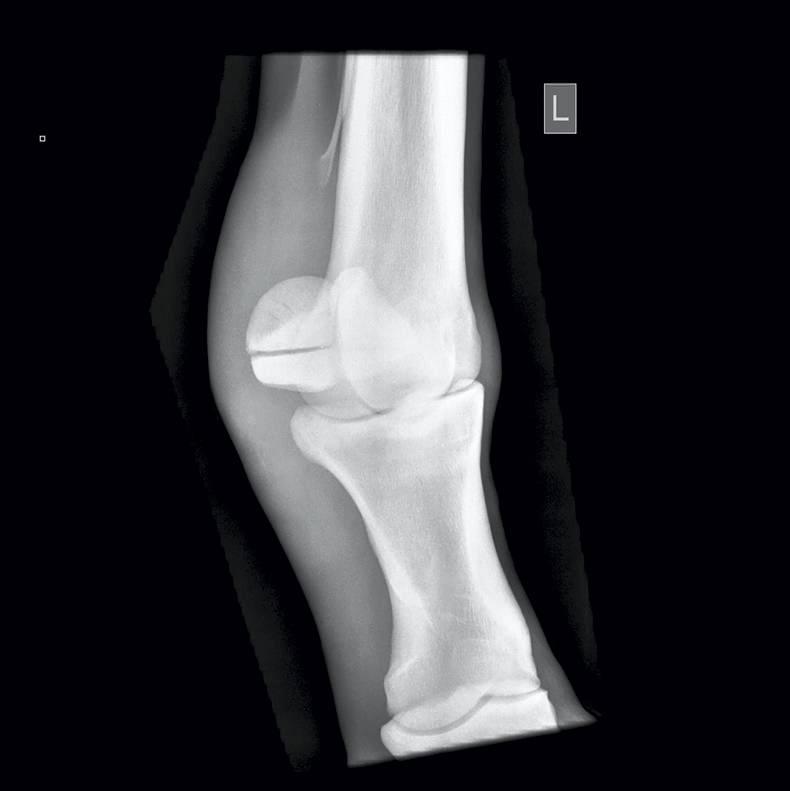
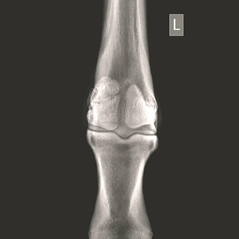
WITH the sales season in full swing, a set of X-rays of a horse involves usually a standard set of 36 views of the fetlocks, knees, hocks and stifles.
A huge number of abnormalities can be found on X-rays, such as sagittal ridge Osteochondritis Dissecans (OCD) defects in the front fetlocks; fragments off the back of the pastern bone; bone cysts in the ulnar carpal bone; and ‘spurring’ in the hock joints.


SHARING OPTIONS: