 This is a subscriber-only article
 It looks like you're browsing in private mode


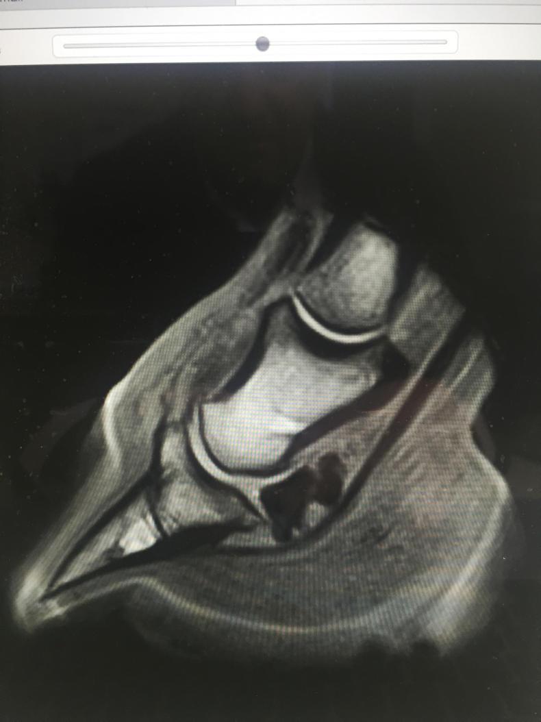
MAGNETIC resonance imaging (MRI) was first performed on live horses at Washington State University in 1997. Over the last 20 years, this diagnostic modality has been intensively developed and improved considerably. Corresponding with the development of the standing MRI scanner, there has been a steep increase in clinical use during the past 10 years. Troytown Grey Abbey Equine Hospital and Hallmarq Veterinary Imaging co-operated to install the only equine MRI unit in Ireland.
Standing equine MRI can image structures along the entire lower limb, from the foot to the knee or hock. Images are taken as slices through the limb, at an angle and position determined by the system operator according to the results of prior tests (such as nerve blocks and X-rays). By positioning images either parallel or at right angles to key structures, any clinical abnormalities such as inflammation, enlargement or degeneration are clearly revealed in a way that has no parallel in X-ray, CT, ultrasound or nuclear scintigraphy (bone scanning).


SHARING OPTIONS: